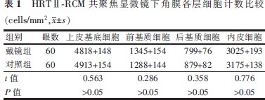
【摘要】 目的 利用激光共聚焦显微镜观察配戴夜戴型角膜塑形镜2年以上患者的角膜组织有无变化。方法 回顾性病例研究。选择2009年1月至2012年12月于南京军区总院眼科就诊且配戴夜戴型角膜塑形镜2年的患者30例(60眼)作为戴镜组,未配戴任何形式角膜接触镜的角膜正常患者30例(60眼)作为对照组。对2组对象进行角膜中央部激光共聚焦显微镜观察(观察组摘镜2~3 h后)。数据采用独立样本t检验进行分析。结果 配戴角膜塑形镜后角膜形态可发生明显变化,上皮至前基质层可见皱褶,上皮下神经丛疏密不均、扭曲,基质神经及内皮细胞形态正常。上皮下神经丛的分布比对照组稀疏,戴镜组的上皮细胞计数为(4818±148)cells/mm2,前基质细胞计数为(1345±154)cells/mm2,后基质细胞计数为(799±76)cells/mm2,内皮细胞计数为(3025±193)cells/mm2,与对照组相比2组差异无统计学意义。结论 长期配戴夜戴型角膜塑形镜对上皮下神经丛数量及眼表形态有影响,对内皮细胞及基质细胞无明显影响。
【关键词】 角膜塑形术; 角膜; 显微镜检查,共焦
DOI:10.3760/cma.j.issn.1674-845X.2014.02.005
作者单位:210002 南京军区南京眼科
通信作者:杨丽萍
Email:1435545746@qq.com
Two-year follow-up of corneal changes from orthokeratology night lens wear using laser confocal microscopy
Xia Yuan, Wang Chunhong, Yang Liping. Department of Ophthalmology, Nanjing General Hospital of Nanjing Military Command, PLA, Nanjing 210002, China
Corresponding author:Yang Liping,Email:1435545746@qq.com
【Abstract】 Objective A laser confocal microscope was used to investigate changes in the cornea from wearing orthokeratology lenses during the night. Methods In a retrospective case study, 60 people were chosen from patients at the Ophthalmology Department of the General Hospital of Nanjing Military Command during January 2009 to December 2012. Among them, 30 patients (total 60 eyes), the experimental group, had worn orthokeratology night lenses for 2 years and the other 30 patients (total 60 eyes), the control group, had never worn any form of orthokeratology. All patients underwent laser confocal microscopy examinations at the centre of the cornea, 2-3 hours after the experimental group had removed the orthokeratology lenses. An independent t test was used. Results Corneal surface shape may change significantly. Wrinkles could be observed from the epithelium to the anterior stroma. The subepithelial plexus was distorted and uneven in density but the stromal nerve and endothelial cell morphology were normal. The epithelial cell count was 4818±148, former stromal cells count was 1345±154, the rear stromal cell count was 799±76, and the endothelial cell count was 3025±193. All these counts were not statistically different from the control group. Conclusion Long-term orthokeratology lens wear during the night can induce changes in the corneal subepithelial plexus but there were no significant changes in stromal or endothelial cells.
【Key words】 Orthokeratology; Cornea; Microscopy,confocal
