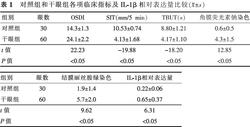
【摘要】 目的 探讨白介素1β(IL-1β)在干眼患者眼表的表达,及其与干眼症状、体征的相关性,阐明IL-1β在干眼发病中的作用。方法 临床试验研究。选取2012年9月至2013年2月来山西就诊的干眼患者30例(60眼,干眼组),无干眼症状体征且年龄与性别构成匹配的个体15例(30眼)作为对照组。所有受检者均做如下检测:眼表失衡指数(OSDI)干眼调查问卷、泪液分泌试验(SIT)、泪膜破裂时间(BUT)、角膜荧光素钠染色、结膜丽丝胺绿染色。同时应用印迹细胞法收集所有2组对象球结膜上皮细胞,分别进行免疫组化染色,以及实时PCR检测结膜细胞中IL-1β的基因表达。采用独立样本t检验,Spearman相关进行数据分析。结果 干眼组与对照组OSDI评分值分别为24.1±2.2、14.3±1.3;SIT分别为(4.13±1.68)mm、(10.53±0.74)mm;BUT分别为(4.17±1.10)s、(8.80±1.21)s;角膜荧光素钠染色评分值分别为4.3±1.5、0.6±0.5;结膜丽丝胺绿染色评分值分别为5.7±2.0、1.9±1.4。干眼组与对照组以上5个指标比较差异均有统计学意义(t=22.23、-19.88、-18.20、12.85、9.62,P<0.05)。干眼组IL-1β相对基因含量为0.65±0.37,对照组为0.22±0.06,二者比较差异有统计学意义(t=6.31,P<0.05)。干眼组IL-1β在结膜上皮细胞中的表达与OSDI评分、角膜荧光素钠染色评分、结膜丽丝胺绿染色评分呈正相关(r=0.81、0.58、0.48,P<0.05),与SIT、BUT结果呈负相关(r=-0.43、-0.45,P<0.05)。通过免疫组化法染色,在干眼患者结膜细胞中可见棕褐色颗粒,而在正常人群中未见棕褐色颗粒。结论 IL-1β作为炎性介质不仅参与了干眼的发病,与干眼的严重程度相关,同时也引起了眼表的损害。
【关键词】 白细胞介素1β; 干眼病; 印迹细胞; 实时聚合酶链反应
DOI:10.3760/cma.j.issn.1674-845X.2014.04.009
基金项目:国家自然科学基金面上项目(81070712)
作者单位:030001 太原,山西(高雅);030002 太原,山西(李冰、陈研遐、常静、郑晓汾)
通信作者:郑晓汾,Email:zxffd@163.com
A study of the expression of IL-1 beta on the ocular surface in dry eye patients
Gao Ya*, Li Bing, Chen Yanxia, Chang Jing, Zheng Xiaofen. * Shanxi Medical University, Taiyuan 030001, China
Corresponding author:Zheng Xiaofen,Email:zxffd@163.com
【Abstract】 Objective To study the expression of IL-1 beta on the ocular surface of dry eye patients and to study its correlation with symptoms and signs of dry eye; to investigate IL-1 beta′s effect on dry eye pathogenesis. Methods Clinical trials. In this case-control study, 30 dry eye patients (60 eyes) and 15 age- and gender-matched control subjects (30 eyes) without symptoms or signs of dry eye were selected from patients who had been treated in our hospital from September 2012 to February 2013. All subjects underwent full ophthalmologic examinations (including OSDI questionnaire, Schirmer′s test (SIT), tear break-up time (BUT), corneal fluorescence staining and conjunctival lissamine green staining). Impression cytology was used to collect conjunctiva epithelial cells from dry eye patients and controls, then these samples were stained by IHC reaction to detect the expression of IL-1 beta by real-time PCR. Results The average OSDI questionnaire scores for the case group and controls were 24.1±2.2 and 14.3±1.3. SIT scores were 4.13±1.68 mm and 10.53±0.74 mm. BUT times were 4.17±1.10 s and 8.80±1.21 s. Corneal fluorescence staining results were 4.3±1.5 and 0.6±0.5. Conjunctival lissamine green staining results were 5.7±2.0 and 1.9±1.4. There were significant differences between dry eye patients and control subjects (t=22.23, -19.88, -18.20, 12.85, 9.62, P<0.05). The average expression of IL-1 beta in the case group and controls was 0.65±0.37 and 0.22±0.06. There were significant differences between dry eye patients and control subjects (t=6.31, P<0.05). The expression of IL-1 beta was positively correlated with scores on the OSDI questionnaire (r=0.81, P<0.05), corneal fluorescence staining (r=0.58, P<0.05), and conjunctival lissamine green staining (r=0.48, P<0.05), and was negatively correlated with BUT (r=-0.45, P<0.05) and SIT (r=-0.43, P<0.05). With IHC staining, brown particles could be seen in the conjunctiva cells in patients with dry eye, but not in the controls. Conclusion IL-1β as an inflammatory mediator is involved not only in dry eye disease, but is also associated with the severity of dry eye and damage on the ocular surface.
【Key words】 Interleukin-1 beta; Xerophthalmia; Impression cytology; Real-time polymerase chain reaction
